
Opening—A speculative problem, grounded in now
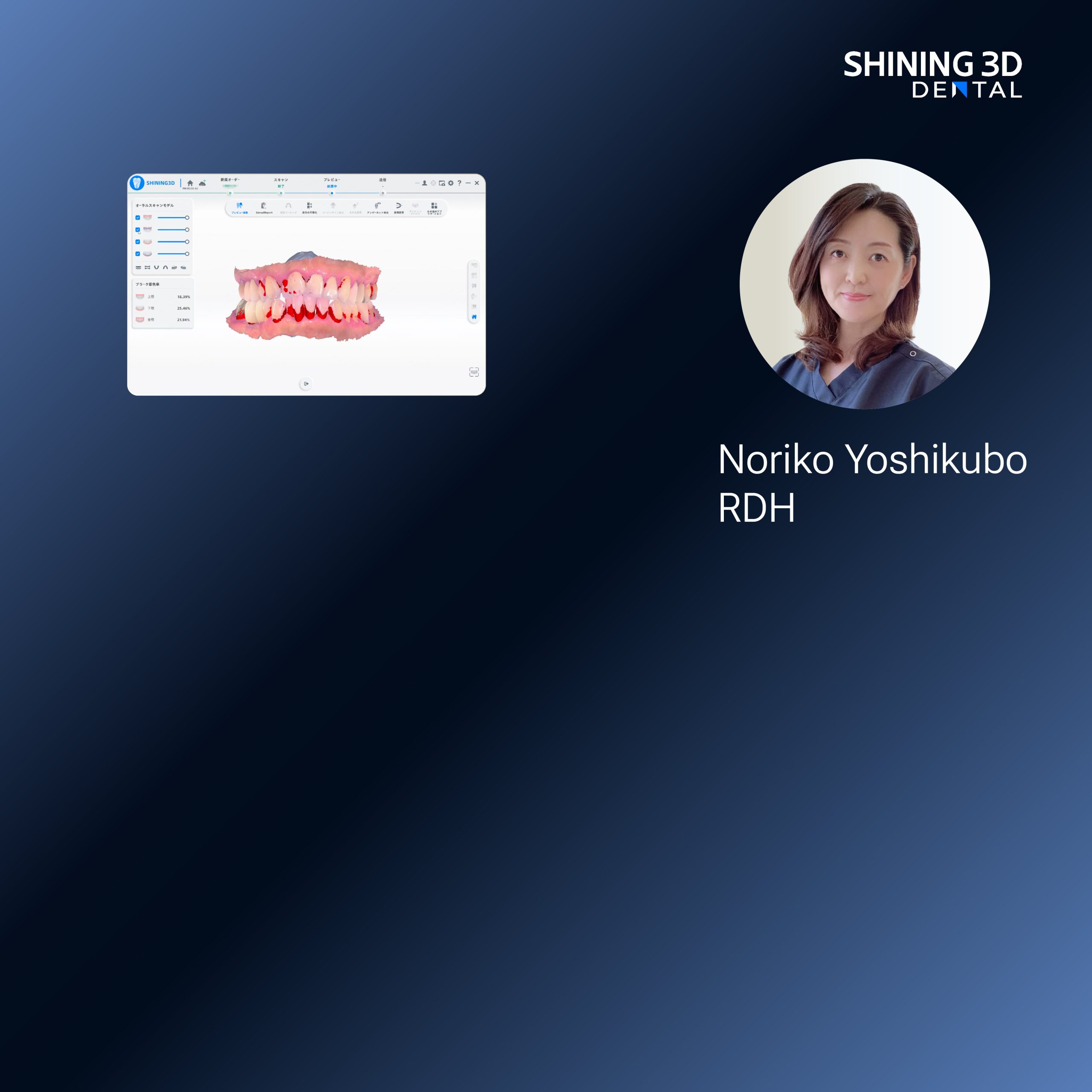
The clinic will look different in ten years: operators co-reading holographic meshes while patients glance at a projected plan. The immediate problem is practical—how do you make today’s scans reliable enough to meet that future? Start with how you capture the mouth. A modern intraoral scanner is a sensor, a light source, and a workflow node; neglect one and the rest fails. At IDS Cologne, public demos showed how crowds watched milling-accurate models generated in seconds by a single dental intraoral scanner, which grounded the speculative promise in an industry-standard moment—practical proof that lighting, posture, and file discipline matter now, not later.
Light: sculpt with controlled illumination
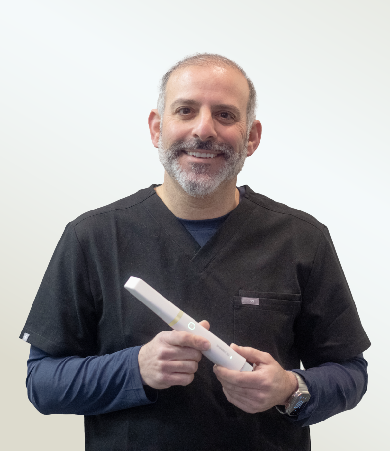
Think of light as the language your sensor reads. Direct, flickering room lamps confuse texture mapping. Use stable, diffuse illumination aimed to minimize reflections on enamel and restorative margins. If the scanner struggles with shiny surfaces, apply a thin, clinically approved scanning spray only where necessary; fewer sprays mean fewer registration artifacts. Watch for mixed lighting—window light plus overhead LEDs produces spectral shifts that look like warp in the final mesh. Solve this by controlling the environment or configuring the scanner’s exposure settings before you scan.
Position: patient, probe, and the choreography of capture
The operator’s hand is a drone in miniature—steady arcs and predictable passes win. Start with the occlusal plane as an anchor line. Approach the arch in consistent, overlapping sweeps: buccal to occlusal to lingual, or the mirrored reverse, keeping the probe perpendicular to the surface when possible. Change posture only to avoid soft-tissue collapse; if the tongue or cheek intrudes, reposition the patient or use retraction. When scanning implants or deep margins, slow your passes and increase overlap so the registration algorithm has abundant common geometry.

File management: formats, fidelity, and traceability
Scanning is metadata as much as mesh. Save original raw captures alongside processed STL exports. Label files with patient ID, date, arch, and scan mode; include a short note on any sprays or retractions used. Use lossless intermediate formats when supported; exporting directly to lossy STL without preserving raw frames discards the data you might need for remeshing. Store backups in two separate locations and keep a version history so you can roll back if the lab requests a different file crop or higher-resolution input.
Common pitfalls and corrective moves
Operator haste creates most errors. Pitfall: scanning too fast across smooth enamel—result is stripe artifacts. Fix: reduce sweep speed and add a deliberate micro-oscillation to give the sensor texture. Pitfall: inconsistent overlap between passes leading to stitching gaps. Fix: plan each arc with at least 30–40% overlap and verify live alignment before proceeding. Pitfall: export that flattens color or loses margin detail. Fix: retain the color/texture layer and export a high-precision mesh for the lab, while giving them a lighter file for preliminary review.
Evidence and authority from practice
I’ve worked alongside prosthodontists and imaging engineers adapting protocols for same-day crowns and multi-unit bridges; consistent outcomes came from three repeatable controls: steady light, deliberate positioning, and documented file workflows. Clinics that adopted these three controls reduced remake requests and shortened lab turnarounds. That practical pattern aligns with what industry demonstrations—like those at IDS Cologne—have shown: robust hardware must be matched with disciplined capture technique to deliver clinical reliability.
Synthesis—where this leads the practice
Speculation about holographic treatment planning becomes useful when you can reproduce a margin three times over and send identical meshes to the lab, the milling center, and archival storage. Control light to reduce noise, control position to preserve geometry, and control files to retain choices and corrections. Those controls are what let a practice scale precise, repeatable outcomes; they’re also the reason clinics choose reliable platforms and partners. For clinics building that capability, SHINING 3D DENTAL represents the practical endpoint of the chain—hardware and workflow designed so the future you imagine starts with every scan you take today.