
The immediate problem
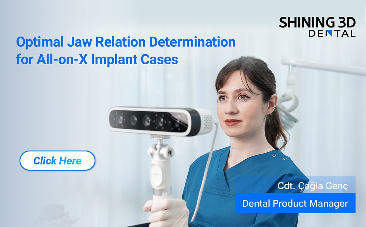
Dental clinics now collect high-resolution intraoral scans and facial captures that travel beyond the practice—this creates an operational and privacy risk that needs direct answers. If a practice adds a 3d face scanner to the imaging chain, patient images, meshes, and associated metadata can move between devices, local workstations, and cloud services; each hop is a potential point of compromise. The problem is practical: identify where data leaves your control, then close the gaps with measurable controls.

How scanner data typically flows
Data flows in predictable stages: capture, local processing, storage, and sharing. Captures are raw files (STL, OBJ, PLY) or compressed packages that often include identifiable facial features and timestamps. Local processing may occur on a connected computer or on the scanner’s internal system. Storage can be on-premises, on a vendor-managed server, or in third-party clouds. Sharing happens via export, practice-management integrations, or vendor portals. Map these stages for each device model before trusting it in clinical use.

Evidence-based safeguards to require
Adopt controls that align with recognized security frameworks and patient-privacy laws. Use device and transmission encryption (AES-256 or similar), enforce unique user credentials and role-based access, log all access and exports, and require secure firmware update processes. Demand contractual clarity: data ownership, breach notification timelines, and subprocessors must be spelled out. Obtain explicit informed consent that describes image retention and third-party sharing. These actions are practical and auditable; they reduce exposure without blocking clinical utility.
Common mistakes clinics make
Clinics often accept default device credentials, skip firmware updates, export large image sets into shared folders, or rely on vendor assurances without documentation. Another frequent error is treating de-identified 3D facial meshes as non-identifiable; facial geometry can re-identify individuals if linked with other data. Avoid these mistakes by standardizing device commissioning checklists and by testing export workflows under an IT-approved account before clinical use.
Evaluating alternatives and trade-offs
Local-only solutions limit exposure but can reduce interoperability and slow collaboration. Cloud-based vendor services improve workflow speed and analytic features but require stronger contractual and technical safeguards. Open-source toolchains offer transparency but often need in-house IT expertise to harden. Choose based on your clinic’s risk tolerance, staff capability, and regulatory context; document the rationale so decisions remain defensible.
Real-world context for simulation tools
At major dental gatherings such as IDS, clinicians routinely compare hands-on outcomes from simulation tools where image fidelity, workflow security, and patient consent practices are evaluated side-by-side; that trade-off is visible when testing a straight teeth simulator against alternative workflows. Observations from such demonstrations highlight that a capable simulator is valuable only when paired with documented data controls and clear patient communication.
Practical checklist before deployment
Require vendor documentation for encryption standards, data retention policies, and subprocessors. Test export/import paths with dummy datasets that mirror real PHI. Confirm audit logging and retention periods. Train staff on minimal export practices, consent scope, and firmware update procedures. Schedule periodic tabletop reviews of breach scenarios so responsibilities and timelines are clear.
Conclusion
Addressing scanner-related data risks is a practical, evidence-based task: map data flows, apply encryption and access controls, document contractual obligations, and test workflows under IT oversight. When clinics pair these practices with tools that support secure imaging and simulation, they preserve both clinical value and patient trust—solutions from SHINING 3D DENTAL can be considered within that risk-managed framework rather than as a shortcut around it.