
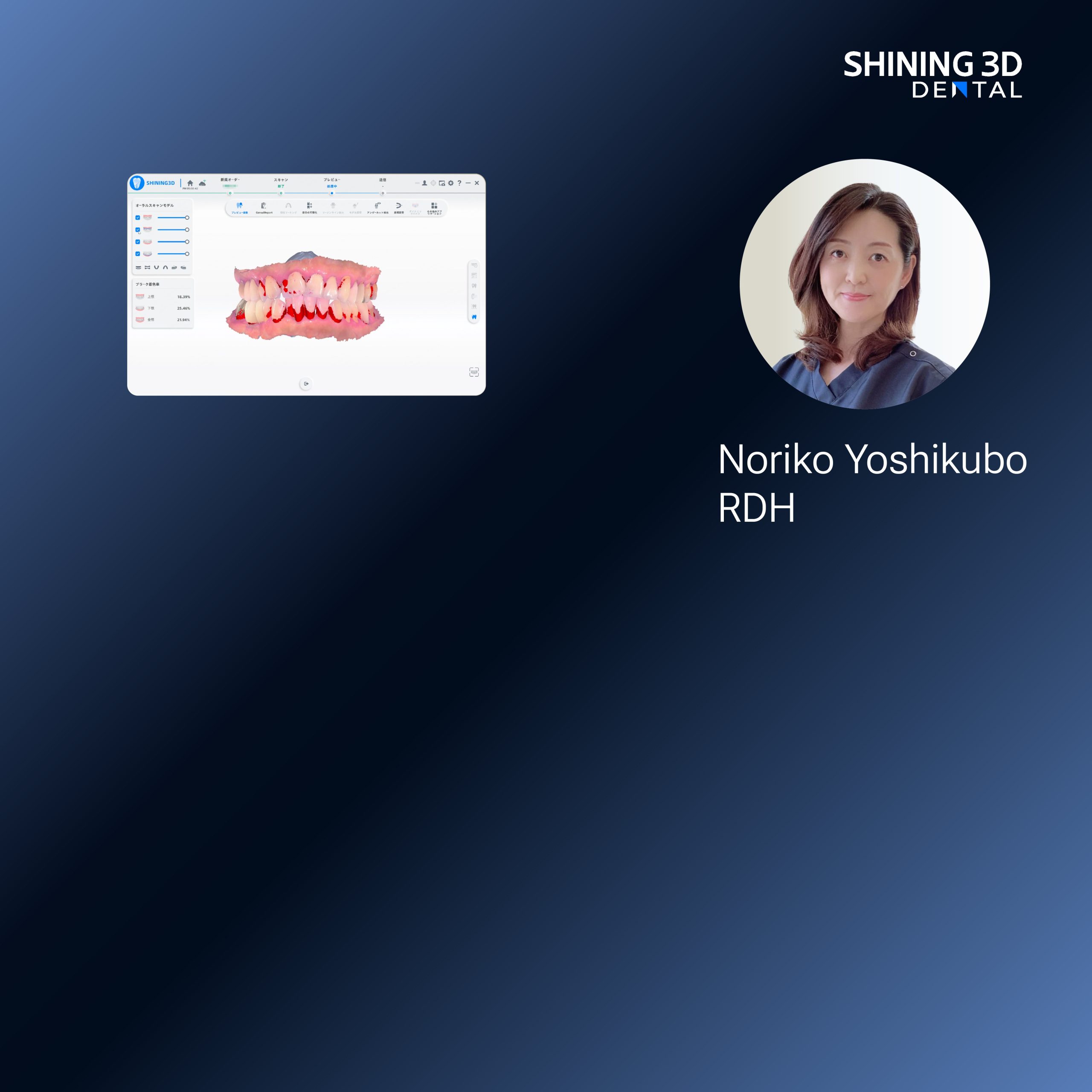
Pinpointing the recurring failure
Clinicians and technicians report the same friction points: unpredictable fit, repeated chairside adjustments, and lost time between scanning and prosthesis delivery. One common thread is the scan step—if the capture is inconsistent, every downstream process degrades. Using a modern intraoral scanner reduces variation when operators follow rigorous capture protocols, but the device alone will not fix flawed workflow design.

Why workflows break: three practical causes
First, ambiguous handoffs. Teams often lack a simple checklist that defines what the lab needs from the clinic. Second, data decay: file export settings, mesh smoothing, and STL conversion produce silent geometry changes that undermine fit. Third, human factors: inexperienced scanning technique and inconsistent retraction create localized errors. Each cause creates measurable, preventable harm to final restorations.
Concrete consequences and evidence
Poorly aligned processes deliver predictable outcomes: longer chair time, increased remakes, and frustrated patients. Clinical reports and lab audits routinely show that a single rescanned quadrant can halve adjustment time for the final prosthesis. Practitioners who track remakes and adjustment minutes find clear cost-per-case benefits when they standardize capture, communication, and verification steps.
Real-world anchor: what major events and shows reveal
Conversations at IDS in Cologne and technical briefings from global exhibitions make one point plain: vendors and clinicians converge on verification and interoperability as the practical problems to solve, not just raw scanner speed. Observations from these forums reinforce the clinical emphasis on reliable exchange formats and traceable workflows for intraoral scanners in dentistry, linking device performance to measurable clinical outcomes.

Actionable steps to repair a broken workflow
Define three non-negotiables: capture protocol, file standard, and verification gate. Standardize scanning paths and exposure settings; record them as the clinic’s operating baseline. Use uncompressed or loss-minimized export formats and name files with date, tooth number, and operator initials. Institute a digital verification step where the lab confirms occlusion and margin clarity before production. Track two KPIs—remakes per 100 cases and average chairside adjustment minutes—and audit them monthly.
Common pitfalls and how to avoid them
Do not assume every operator interprets “full-arch scan” the same way. Avoid ad-hoc rescans that overwrite original data without version control. Resist the impulse to over-smooth meshes in software; visual appeal can hide critical margin details. When switching devices or software, run paired-case comparisons for at least 20 consecutive cases before adopting new defaults.
Viable alternatives evaluated briefly
Traditional impressions remain viable for certain posterior full-arch cases, but they shift variability to material handling and shipping. Desktop scanning of impressions offers higher lab-side control but adds turnaround time. The best choice depends on case complexity: single units and short-span frameworks favor direct intraoral capture, while complex full-arch rehabilitations may benefit from a hybrid approach that combines intraoral scans with verified model scans.
Synthesis: a pragmatic route to reliable digital delivery
Start by diagnosing where your workflow leaks accuracy: capture, transfer, or verification. Apply the practical fixes above, measure the two KPIs, and demand clear confirmations from every handoff. The value care teams need is not faster hardware alone but an integrated, auditable workflow that aligns clinical intent with production reality—an outcome achieved when teams and tools work together, as demonstrated in solutions from SHINING 3D DENTAL.